Request a quote
Medical

IKEM prints 4 to 6 kidneys a month before transplant surgery

A Prague transplant institute prints full-scale organ models from CT data so surgeons can find the complications before the operation starts.

4 to 6
models a month
12 to 18 h
print time each
2
printers
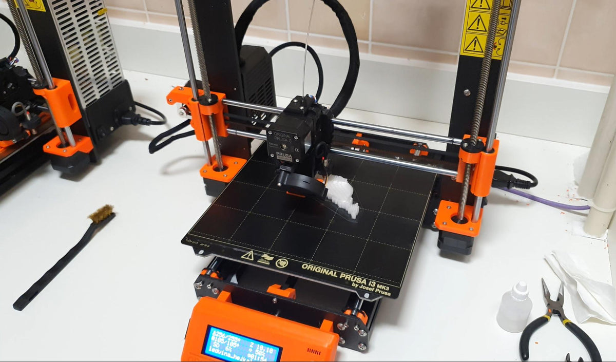
An Original Prusa i3 MK3 on a worktop in front of a tiled wall, printing the first white layers of a kidney model on its black build sheet, with a second MK3 standing at the left edge of the frame.
An Original Prusa i3 MK3 at IKEM, printing the first layers of a kidney model.

In the write-up:

  • Two previous-generation Prusa printers doing the work
  • Four to six kidney models printed per month
  • 12 to 18 hours of print time per kidney
  • Models printed in ABS at real-life scale, not reduced
  • Workflow: CT scan, segmentation, conversion to a 3D object, slicing, print
  • Aorta models printed from blood-vessel segmentation are used to rehearse catheter insertion
  • Donors are given their printed organ model after the operation

What they print. The Institute for Clinical and Experimental Medicine in Prague, IKEM, prints models of organs while preparing for transplant surgery. Four to six kidneys a month come off two previous-generation Prusa printers, in ABS, at real-life scale. Each one takes 12 to 18 hours.

The workflow. A CT scan captures the organ in place. Segmentation joins the separate image slices into one volume, specialist software converts that to a 3D object, and a slicer prepares the file. Nothing about the chain is exotic; the constraint is the segmentation work, not the printing.

Why a model and not a screen. Surgeons can examine the organ physically before the operation begins and spot possible complications in advance. The institute is explicit that being able to hold the organ, rather than rotate it on a monitor, is what made the models useful in practice. The same approach extends to vasculature: segmenting the blood supply gives an aorta model that catheter insertion can be tried on first.

Who else uses them. Medical students learn on the models, and they are used to explain the procedure to patients, which is a job a scan cannot do. After the operation the donor is given their printed organ as a keepsake.

How it started. Milan Bergman, the application manager who set it up, described 3D printing as completely new technology for the institute that the team got a grasp of quickly. That is the realistic version of a hospital printing program: two desktop machines, a handful of models a month, and a workflow built around imaging data the hospital already has.

Why the scale is modest. Four to six models a month on two printers is not a capacity limit. A kidney occupies a machine for 12 to 18 hours, so two printers cover well over the current caseload; the bottleneck is the imaging and segmentation work in front of the printer, and the surgical calendar behind it. A hospital starting this does not need a fleet, it needs one person who can segment a scan reliably.

What it is not. These are planning and teaching models, not implants. The printed organ is a reference object a surgeon handles before an operation, printed in ABS on a standard desktop machine. That is the whole reason it is affordable enough to do routinely for four to six cases a month.

Setting up something similar? Talk to an expert or see our business, education and government sales.

Summarized from Prusa Research's story on the Institute for Clinical and Experimental Medicine (blog.prusa3d.com, 2019).

Planning a fleet of your own?

Send us your monthly volumes and we will size the fleet, the power and the spares list with you.

Request a quote
Or call (302) 439-0098

Running Prusa hardware in production?

Tell us what you print and how many machines it takes. You approve every word before it goes live.

Send it to us